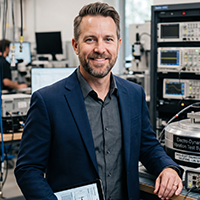
The Focused Ion Beam in dual beam configuration with Scanning Electron Microscope (FIB-SEM) is the most advanced electron microscope apparatus within the section. This system combines the use of the electron beam for imaging and analysis with an additional beam administering a high current of gallium ions (Ga+). This arrangement allows for an extension of analytical capabilities.
Principle and Methodology of FIB-SEM
The focused ion beam-scanning electron microscope (FIB-SEM) uses a second beam, the ion beam, to cut through the material while the SEM does high-resolution imaging. This method can create a 3D stack of data that represents your sample layer by layer at the nanometer scale.
Sample preparation involves mounting samples on a holder and coating them with a conductive layer. Scanning Electron Microscope imaging uses electron beam scanning to obtain high-resolution images. FIB milling creates defined areas of the sample. Compounding sequential FIB milling and SEM imaging allows for 3D reconstruction.
Instrumentation of FIB-SEM
This system is a multi-component system having an electron column for SEM imaging, an ion column for FIB milling, detectors to capture signals, a sample stage for precise positioning and movement, a Gas Injection System (GIS) for enhanced material deposition, and control software for beam control, image acquisition, and data processing.

Applications of Focused Ion Beam Scanning Electron Microscope
This is also easily utilized to prepare TEM samples in the study of extraterrestrial materials, such as presolar grains, high-pressure mineral phases, and other meteorite components. Focused Ion Beam Scanning Electron Microscope is used in various industries, including Materials Science, Semiconductor Industry, Nanotechnology, Biological Research, and Geosciences.