ESEM was developed to overcome the limitations of conventional SEM. With its specialized electron detectors and differential pumping systems, which allow the electron beam to be transferred from the high vacuum in the gun area to the high pressure achievable in its specimen chamber, the ESEM is a complete and unique instrument designed for imaging specimens in their natural state.
Principle and Methodology of ESEM
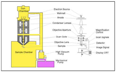
An ESEM uses a scanned electron beam and electromagnetic lenses to focus and direct the beam onto the specimen surface in the same way that a traditional SEM does. A very small, focused electron spot (probe) is scanned in raster form across a small specimen region. The beam electrons interact with the specimen surface layer, producing a variety of signals (information) that can be gathered using appropriate detectors. The output of these detectors modulates the screen of a monitor, using appropriate electronics, to generate an image that corresponds to the small raster and information originating from the specimen surface, pixel by pixel.
Instrumentation
The significant components of ESEM are the electron source to provide a focused electron beam, differential pumping system to maintain pressure gradients between the high-vacuum electron column and the low-vacuum specimen chamber, specimen chamber to accommodate varying pressure levels, and the introduction of gases, detectors, control and imaging system that include both software and hardware to control the microscope parameters and to capture and process images.
Applications of ESEM
ESEM is a versatile imaging technique used in different fields, including biological sciences, materials science, industrial applications, geology, and food science. It permits the observation of non-conductive and hydrated specimens, providing an understanding of cell structure and function without dehydration or coating. It is also used in materials science to study properties of materials sensitive to high vacuum and environmental conditions.